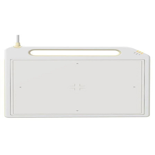
Product Description
Dr. Tech X-Ray Detector measures incoming X-rays using an electric current. An electron is released in this sort of detector when an X-ray interacts with a substance. In the detector, that electron may jiggle around and excite more electrons. Processing software is used to create the finished image. Our provided Dr. Tech X-Ray Detector is used to directly acquire x-ray pictures in place of film or computed radiography devices. This must be used to depict the electrical charge that was converted from absorbed X-ray energy.
| Greyscale |
16 Bits |
| Pixel Area |
434 x 355 mm 2 |
| Pixel Array |
2816 x 2304 |
| Pixel Pitch |
154 um |
| Sensitivity |
0.62 CT/Ngy |
| Size |
14" x 17" |
| Weight |
4 kg |
High-Quality Imaging PerformanceEquipped with a pixel size of 140 m and a resolution of 3.5 lp/mm, the DR Tech X Ray Detector ensures exceptional image accuracy for a variety of diagnostic and inspection applications. Its advanced scintillator options (CsI/GOS) deliver superior sensitivity and clarity for both standard and complex imaging procedures.
Versatile Applications and UsageSuitable for medical, clinical, veterinary, and non-destructive testing environments, this detector supports both cable and wireless connectivity. Its automation grade, semi-automatic operation, and compatibility with PC or workstation displays make it ideal for high-throughput and portable imaging needs.
Robust Design and Long-Lasting ReliabilityEngineered to withstand over 100,000 exposures, the detector integrates a built-in Li-Ion battery for wireless models and is protected with an IPX4 waterproof rating. Its lightweight design (approx. 4.5 kg) and standard detector connector facilitate seamless system integration and mobile workflows.
FAQ's of DR Tech X Ray Detector:
Q: How is the DR Tech X Ray Detector operated and controlled?
A: The detector supports software and PC control modes, allowing users to manage imaging processes directly from a connected computer workstation. Automated exposure detection further streamlines efficient imaging workflows.
Q: What are the benefits of its wireless capability and built-in battery backup?
A: The wireless models incorporate a built-in Li-Ion battery, enabling cable-free operation and flexible positioning. This enhances portability, facilitates quick relocation, and ensures uninterrupted imaging even during power fluctuations.
Q: When is DICOM 3.0 compatibility important for this device?
A: DICOM 3.0 compatibility is crucial for integrating the detector into hospital and clinical networks, ensuring seamless data management, image sharing, and compliance with standardized medical imaging protocols.
Q: Where can the detector be installed or mounted?
A: The detector is designed for versatile mounting options, including tabletop, wall-mount, and portable setups. Its compact size and robust construction allow it to be utilized in a range of clinical, laboratory, and on-field environments.
Q: What process does the device follow for image acquisition and transfer?
A: After exposure, the detector converts X-ray signals to high-resolution digital images within less than 5 seconds. Images are then transmitted to PC workstations via Gigabit Ethernet or optional Wi-Fi, supporting efficient workflow and rapid review.
Q: How does the detector ensure longevity and reliable performance?
A: Designed for over 100,000 exposures, the detector employs durable hardware, an IPX4 waterproof rating, and robust scintillator materials, guaranteeing consistent performance in demanding clinical and industrial settings.